Blood Flow to Little Toe Uses Which Vein
Veins carry blood back toward your heart. This is best considered when the patient is lying down.
The Venous System Of The Foot Anatomy Physiology And Clinical Aspects Servier Phlebolymphologyservier Phlebolymphology
There are three types of blood vessels.

. Arteries carry blood away from your heart. As your muscles contract and relax they squeeze around the large veins in your legs promoting healthy circulation in your lower body. Tingling in toes as well as the sensation of the foot.
There are two systolic velocities S 1 and S 2 and two diastolic velocities D in early diastole and atrial reversal flow Ar in late diastole the latter being a result of atrial contraction. Blood flows back to the chest from the legs through the veins. Its often used to detect narrowing of the carotid arteries or to determine if you have plaque buildup causing carotid disease hardening of the arteries.
It is accompanied by the. Walking is the best way to quickly improve circulation. Your body contains about 60000 miles of blood vessels.
A protocol for using finger-toe pulse oximetry so attaining the Lanarkshire oximetry index to determine whether a patient is suitable for compression therapy is outlined below Bianchi 2005. Too little blood flow ischemia results in tissue death. The Descending Thoratic Aorta splits into some arteries which.
The left ventricle pumps the blood through the Descending Thoratic Aorta get the blood to your legs and feet - where the toes are. If sitting for a long period. Capillaries the smallest blood vessels connect arteries and veins.
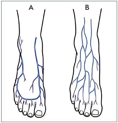
The superior surface of the foot drains into the digital veins and the inferior surface drains into the plantar veins which flow into a complex series of anastomoses in the feet and ankles including the dorsal venous arch and the plantar venous arch. - Place the pulse oximeter sensor on any finger Fig 1a. The abdominal aorta forms several branches three of which supply blood to the intestines.
Once the blood has travelled through the arteries into the capillaries which lie in all of the tissues of the body the. This occurs each time you take a step as your medial longitudinal arch stretches and pronates followed by contraction and flexion. Decreased Pressure on the Posterior Tibial Artery Vein.
Palpable over the dorsum of the foot just lateral to the extensor hallicus longus tendon. This region is a significant route for lower leg. A painless noninvasive test that uses sound waves to create pictures of the carotid arteries in the neck.
Thats because every step you take helps activate the muscles in your legs which helps increase blood flow. In a normal person the heart pumps arterial blood that is full of oxygen and nutrients out into the body limbs and head through the arterial system. Medial plantar artery.
From the dorsal venous arch blood supply drains into the anterior and. It is for these 2 reasons that veins do not Pulsate which is why you cant feel a pulse in the veins only in the arteries. Cleveland Clinic is a.
It is accompanied by the dorsalis pedis vein. Too much blood can raise intracranial pressure ICP which can compress and damage delicate brain tissue. Splits into capillaries the smallest type of blood vessel which gives the blood to your toe.
As we have said the blood is not being pushed back by the heart and so it has Low Pressure and fairly smooth Flow when you are lying down at rest. The celiac trunk superior mesenteric artery and inferior mesenteric artery. The stretching regime made arteries less stiff which helped them dilate.
Arcuate see below lateral tarsal. From the dorsal venous arch blood supply drains into the anterior and posterior tibial veins. Anterior lateral malleolar artery.
CBF is tightly regulated to meet the brains metabolic demands. Found that performing simple leg stretches can help improve vascular function after 12 weeks. While at rest or when walking between step propulsion and when the heel touches down blood is pooled in the foot veins.
The dorsal digital veins from the toes take this deoxygenated blood into the dorsal venous arch. - Ensure heshe is lying comfortably in a semi-recumbent position. The posterior tibial artery and vein are key players in foot circulation.
When blood returns to your heart it flows to your lungs to receive oxygen. Supplies the medial side of the foot abductor hallucis muscle and flexor digitorum brevis muscle. This artery supplies blood to the surface of the foot as a continuation of the anterior tibial artery.
Correct Toes helps open up that space between the toes so that more blood can actually reach the toe tissues. Then your heart pumps that blood out to the rest of your body and the process begins again. They then merge with the posterior tibial veins and form the popliteal vein situated posterior to the knee.
Oxygenated blood leaves the heart through the aorta which descends into the abdominal cavity as the abdominal aorta. This then connects with the anterior tibial veins which drain the anterior leg compartment and foot Uhl et al 2017. Gives off cutaneous branches that perforate the plantar aponeurosis between abductor hallucis muscle and.
Feel free to review the other videos in the playlist at. Anterior medial malleolar artery. In an adult CBF is typically 750 milliters per minute or 15 of the cardiac output.
Provides the arterial digital supply to the big toe. Nerves that are required for muscle movement as well as those that are responsible for the control of heart rate and blood pressure can be affected in peripheral neuropathy. Branch of the posterior tibial artery.
The superior surface of the foot drains into the digital veins and the inferior surface drains into the plantar veins which flow into a complex series of anastomoses in the feet and ankles including the dorsal venous arch and the plantar venous arch Figure 20520. Pulmonary vein flow when added to the mitral inflow velocity is very helpful in the assessment of diastolic function. Imaging tests that provide detailed information about blood vessels.
A continuation of the anterior tibial artery in the foot. To use a rough analogy what happens to these blood vessels is akin to a garden hose that gets slightly kinked. In the foot the plantar venous plexus pump initiates venous return.
The posterior tibial artery. Each of these arteries forms many smaller branches that. This video is part of a playlist on blood vessel anatomy physiology at my youtube channel drjahn41.
Blood flows through a network of vessels called the circulatory system. As a result in addition to poor circulation in toes other symptoms may be present as well such as pain and numbness.

Foot Blood Flow Images Stock Photos Vectors Shutterstock
Pin On Natural Remedies
Signs And Symptoms Of Poor Circulation In Feet Advanced Foot Ankle Care Specialists

Vascular Changes Associated With Ageing Erica Dash Podiatry
No comments for "Blood Flow to Little Toe Uses Which Vein"
Post a Comment